PhysicalTherapy.com Phone: 866-782-6258


Management of the Elbow Complex

Management of the Elbow Complex
David Nolan, PT, DPT, MS, OCS, SCS, CSCS
November 17, 2016
Share:

Learning Objectives

  • Accurately describe at least two examples of the anatomic and bow mechanical relationships of the elbows that relate to normal function.
  • Describe without queues the pathomechanics of at least two selected elbow injuries.
  • Independently outline an evidenced based examination with at least three intervention techniques for selected elbow conditions.

Anatomy

Articulations

There are three primary articulations: the humeral ulnar, humeral radial, and the proximal radial ulnar joint.

Carrying Angle

Another thing to consider when reviewing anatomy is the carrying angle.  There is a normal valgus angulation. In men, that normal angulation ranges between 11 and 14 degrees, and in women, 13 to 16 degrees. The female pelvis is a bit wider which may be a component on why there is a difference in gender. Again, keep in mind the normal position of the elbow is going to have a bit of a valgus angulation.

Ligamentous Complex

Medial ligament complex. The ulnar collateral ligament complex is found on the medial side of the elbow.  It has three different bundles.  The anterior bundle is going to be taut in extension, the posterior bundle will be taut in flexion, and there is also a transverse bundle. Thickenings of the structure provides some variability in the direction of the fibers, which is going to be more helpful as far as trying to control load.

Restraint to Valgus Stress

Specific literature provides us information regarding restraint to valgus stress and how much each component is contributing.  The joint surfaces, or joint articulation will give us some stability, as well as the ligamentous structures and the capsule.  How much each is contributing differs a little bit depending on whether or not the elbow is flexed versus extended. You can see those percentages in Figure 1. The joint articulation is giving between 30 and 35% restraint both in elbow flexion and extension. The ligaments are providing a more significant barrier in elbow flexion compared to elbow extension, where as the joint capsule is providing much more restraint in elbow extension compared to elbow flexion.

Table of restraints to valgus stress with elbow extension and elbow flexion values noted for joint articulation medial ligaments and joint capsules

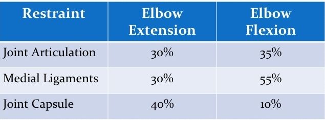
Figure 1.  Restraints to Valgus Stress

Lateral Ligament Complex

Looking laterally at the lateral ligament complex:

  • Radial Collateral Ligament: taut throughout the entire range of motion.
     
  • Lateral Ulnar Collateral Ligament: primary restraint to varus stress at the elbow.
     
  • Accessory Collateral Ligament: fibers will blend in with the annular ligament.
     
  • Annular Ligament: helps stabilize the proximal radial ulnar joint. The anterior portion of the annular ligament is taut in supination, and the posterior portion is going to be taut in pronation.                                                                                                                                          

Restraint to Varus Stress

Figure 2 displays the percentages for primary restraint to varus stress.  Joint articulation provides quite a bit of restraint in both elbow extension and flexion. About 55% restraint is happening with joint articulation and elbow extension.  It is as high as 75% when the elbow is flexed. The lateral ligaments and joint capsule are providing about 45% in elbow extension, and only about 10% in elbow flexion. Other soft tissue (the other musculature that's crossing that portion of the joint), doesn't help provide much restraint in elbow extension. Again, elbow extension is going to be our close-packed position, so we're relying on joint articulation, capsular ligamentous structures and extension. There is a small amount of restraint (15%) in elbow flexion.  This is more of an open-packed position from the corresponding soft tissues and musculature around the elbow with regard to varus stress.

Table of restraints to varus stress with elbow extension and elbow flexion values noted for joint articulation lateral ligaments and joint capsules and other soft tissues

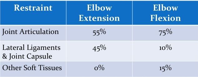
Figure 2. Restraints to Varus Stress


david nolan

David Nolan, PT, DPT, MS, OCS, SCS, CSCS

Dr. Nolan is an Associate Clinical Professor at Northeastern University in the Department of Physical Therapy, Movement and Rehabilitation Sciences as well as a Graduate Lecturer in the College of Professional Studies in the transitional Doctor of Physical Therapy Program at Northeastern University.  David is also a Clinical Specialist at the Mass General Sports Physical Therapy Service and the Director of the MGH / Northeastern University Sports Physical Therapy Residency Program. 

Dr. Nolan is a board certified Orthopedic Clinical Specialist and Sports Clinical Specialist through the American Board of Physical Therapy Specialties and a Certified Strength and Conditioning Specialist through the National Strength and Conditioning Association.  He is a past recipient of the “Excellence in Clinical Teaching” award from the New England Consortium of Academic Coordinators of Clinical Education as well as the award for Outstanding Achievement in Clinical Practice by the Massachusetts Chapter of the APTA.  Dr. Nolan was also presented with the 2019 Lynn Wallace Clinical Educator award from the American Academy of Sports Physical Therapy. 


Related Courses

Management of Adhesive Capsulitis
Presented by David Nolan, PT, DPT, MS, OCS, SCS, CSCS
Recorded Webinar
Course: #4438Level: Intermediate2 Hours
Adhesive capsulitis is characterized by a painful, gradual loss of motion which may lead to significant functional limitations. This session reviews the epidemiology, classification, and differential diagnosis associated with adhesive capsulitis. Current evidence related to conservative physical therapy management utilizing exercise and joint mobilization is reviewed.

Editor's Note: Regarding Pennsylvania credits, this course is approved by the PA State Board of Physical Therapy for 1 hour of general and 1 hour of Direct Access CE credit.

Managing Foot and Ankle Pathology in the Distance Runner
Presented by David Nolan, PT, DPT, MS, OCS, SCS, CSCS
Recorded Webinar
Course: #4455Level: Intermediate2 Hours
Running related injury is a common presentation in physical therapy practice. Some studies report over 50% of runners will experience an injury that limits participation at some point. The majority of injuries specific to distance runners will occur in the lower quarter and many of these will involve the lower leg and foot and ankle complex. This course will review the epidemiology of selected running related injuries and provide evidence-based recommendations for examination and treatment.

Rotator Cuff Lesions: Conservative and Post-Operative Treatment
Presented by David Nolan, PT, DPT, MS, OCS, SCS, CSCS
Recorded Webinar
Course: #3441Level: Intermediate2 Hours
Rotator cuff pathology is common in the general population and even more frequent with overhead athletes and workers. The purpose of this presentation is to present an evidence-based review of current physical therapy practice for the conservative and post-operative management of rotator cuff pathology. This course is directly related to the practice of physical therapy and athletic training and is therefore appropriate for the PT/PTA and AT.

Management of Patellofemoral Pain Syndrome
Presented by David Nolan, PT, DPT, MS, OCS, SCS, CSCS
Recorded Webinar
Course: #3475Level: Intermediate2 Hours
Patellofemoral Pain Syndrome is a common diagnosis for patients presenting to physical therapy with anterior knee pain. The purpose of this presentation is to present an evidence-based review of current physical therapy practice for the management of patellofemoral pain syndrome. Evidence-Based therapeutic exercise interventions will also be reviewed. This course is directly related to the practice of physical therapy and athletic training and is therefore appropriate for the PT/PTA and AT

Complex and Chronic Impairment in Concussion
Presented by Laura Morris, PT, NCS
Recorded Webinar
Course: #4353Level: Intermediate2 Hours
This webinar will include an exploration of the various etiologies of dysfunction following mTBI, including, headache, visual/oculomotor impairment, chronic dizziness, and pain. The challenging issue of prioritizing intervention and appropriate referral to other medical practitioners will be discussed. This course is directly related to the practice of physical therapy and athletic training and is therefore appropriate for the PT/PTA and AT.

Editor's Note: Regarding Pennsylvania credits, this course is approved by the PA State Board of Physical Therapy for .5 hour of general and 1.5 hour of Direct Access CE credit.

Our site uses cookies to improve your experience. By using our site, you agree to our Privacy Policy.